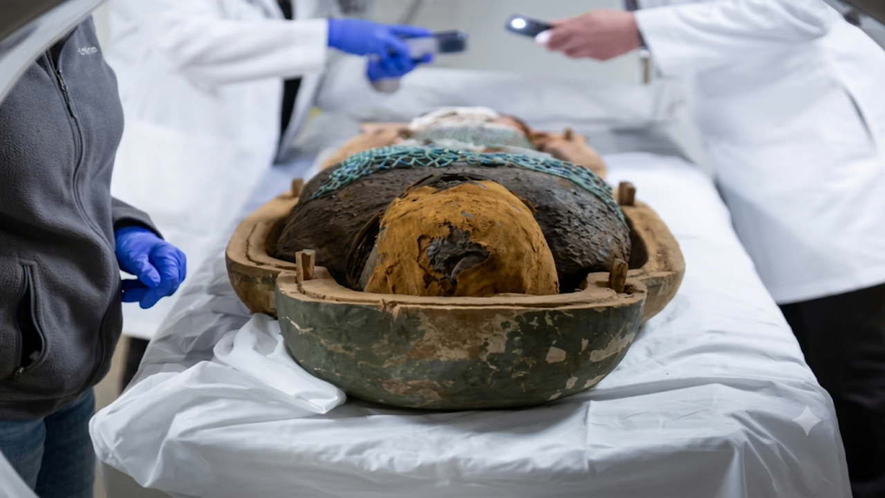
A groundbreaking medical imaging project in Los Angeles has provided researchers with an extraordinary glimpse into the lives of two Egyptian mummies dating back more than 2,200 years. Using state-of-the-art scanning technology capable of capturing images with half-millimeter precision, doctors and scientists conducted detailed examinations of the ancient remains without disturbing their wrappings.
What began as a routine effort to better understand the mummies’ health and preservation soon led to an unexpected discovery. Researchers identified a spinal condition in one of the mummies that challenged assumptions about ancient health and disease, demonstrating how modern medical technology can continue uncovering secrets hidden within archaeological treasures centuries after their burial.
High-Tech Scans Unlock Ancient Secrets
The examination was conducted using advanced CT imaging technology in Los Angeles, allowing specialists to create highly detailed three-dimensional views of the mummies’ internal structures. Unlike traditional archaeological methods that sometimes require unwrapping or physically examining remains, the scanning process enabled researchers to study bones, organs, and preserved tissues while leaving the mummies completely intact.
The high-resolution images provided unprecedented insight into the physical condition of the ancient individuals. Every bone, joint, and anatomical feature could be examined in remarkable detail, helping scientists reconstruct aspects of their lives, health conditions, and even possible causes of death.
Researchers emphasized that modern imaging technology has transformed mummy studies by allowing experts from multiple disciplines—including medicine, anthropology, archaeology, and radiology—to collaborate on investigations that were once impossible. The scans revealed information about age, physical characteristics, and medical conditions that had remained hidden for more than two millennia.
Unexpected Discovery Found in Ancient Spine
While reviewing the images, researchers identified a surprising spinal abnormality in one of the mummies. The finding attracted particular attention because it provided evidence of a condition not commonly documented in ancient remains. The discovery demonstrated that certain degenerative or structural spinal conditions affected individuals thousands of years ago, much as they do today.
Doctors involved in the analysis noted that the precision of the scans allowed them to observe subtle skeletal changes that may have gone unnoticed using older technologies. The spinal condition appeared preserved well enough to enable detailed medical evaluation despite the passage of centuries.
The finding offered researchers an opportunity to compare ancient and modern health conditions. By studying how diseases and skeletal disorders manifested in historical populations, scientists can gain a deeper understanding of human health across different eras and environments. Such discoveries contribute valuable information to both medical science and archaeology.
What Researchers Learned About the Mummies
Beyond the spinal finding, the scans revealed a wealth of information about the two ancient Egyptians. Researchers were able to estimate their ages, examine their bone structure, and study evidence of their lifestyle and overall health. Details about dental health, skeletal development, and possible nutritional conditions emerged from the analysis.
The examinations also provided insights into ancient Egyptian mummification practices. Researchers observed how the bodies had been prepared, preserved, and wrapped before burial. Such information helps historians better understand cultural and religious traditions associated with death and the afterlife in ancient Egypt.
In addition, scientists were able to assess the condition of the mummies without risking damage to the fragile remains. This non-invasive approach has become increasingly important as museums and research institutions seek to balance scientific investigation with preservation of cultural heritage.
Mummy Scan Project Overview
| Category | Details |
|---|---|
| Subjects Studied | Two Egyptian mummies |
| Estimated Age | Approximately 2,200 years old |
| Location of Scan | Los Angeles, California |
| Technology Used | Advanced CT imaging |
| Scan Precision | Half-millimeter resolution |
| Main Discovery | Unexpected spinal condition |
| Research Fields Involved | Medicine, archaeology, anthropology |
| Method | Non-invasive examination |
| Additional Findings | Health, age, and mummification details |
| Historical Significance | New insights into ancient health conditions |
The Role of Modern Medicine in Archaeology
The project highlights the growing partnership between medical science and archaeology. Technologies originally developed for diagnosing living patients are increasingly being used to investigate historical remains and ancient artifacts. CT scanners, MRI systems, and digital reconstruction software have become powerful tools for studying the past.
Medical professionals bring unique expertise to these investigations by identifying diseases, injuries, and anatomical conditions that may not be immediately recognizable to archaeologists. Their involvement helps transform ancient remains into valuable sources of scientific information about historical populations.
Researchers believe that continued advancements in imaging technology will lead to even more discoveries in the future. Improved resolution and artificial intelligence-assisted analysis may reveal details that remain invisible today, opening new possibilities for understanding ancient civilizations.
Why the Discovery Matters
The spinal finding is significant because it demonstrates that health challenges often considered modern may have affected humans for thousands of years. Studying ancient diseases provides a broader perspective on the development of medical conditions and how they have evolved over time.
The discovery also underscores the importance of preserving archaeological remains. Every new technological advancement creates opportunities to revisit ancient specimens and uncover information that previous generations of researchers could not access. As a result, mummies and other historical remains continue to contribute to scientific knowledge long after their discovery.
For museums and educational institutions, findings like these help engage the public by connecting ancient history with modern science. They illustrate how technology can bridge thousands of years of human experience and reveal stories hidden within the past.
The high-precision scanning of two Egyptian mummies in Los Angeles has demonstrated the remarkable potential of modern medical technology in archaeological research. By examining the remains with half-millimeter accuracy, scientists uncovered valuable information about ancient health, preservation practices, and human anatomy.
The unexpected discovery of a spinal condition in one of the mummies highlights how ancient remains can continue providing new scientific insights centuries after their burial. As imaging technology advances, researchers expect even more revelations that will deepen our understanding of ancient civilizations and the people who lived within them.
FAQ’s:
What was the main discovery made during the scans?
Researchers identified an unexpected spinal condition in one of the 2,200-year-old Egyptian mummies.
Where were the scans conducted?
The examinations were performed in Los Angeles using advanced medical imaging technology.
What technology was used?

Scientists used high-resolution CT scanning capable of capturing images with half-millimeter precision.
Why are mummy scans important?
They allow researchers to study ancient remains without damaging them while revealing information about health, age, and cultural practices.
What can scientists learn from ancient medical conditions?
Studying ancient diseases helps researchers better understand the history and evolution of human health across different time periods.